
The first step to ensuring that you protect your vision is having a complete eye examination. How often you have an eye examination depends on your age and general health. As you get older (55+) it is important to have a complete eye examination at least every two years. Your eye doctor will tell you what is best for you. If you are a diabetic, you should have a dilated eye examination every year. We encourage you to call and schedule an appointment at any of our offices throughout Northern Maine.
A complete eye exam usually includes the following:

Additional Testing
In some cases, additional tests are required to fully assess and diagnose eye disease. Common tests include:
Optical Coherence Tomography OCT – This test uses light waves to take cross section pictures of your retina, lens or cornea. It allows the doctor to examine the layers of each part of the eye to identify abnormalities such as macular degeneration.
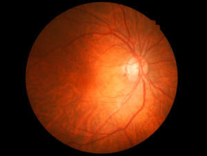
Fundus Photos – these high resolution photographs of the retina help identify and document the presence of certain eye diseases like diabetic retinopathy. Photographs are used to compare the progression of eye disease from one visit to the next. This help the doctor see how well a particular treatment is working.
Visual Field – This test measure how well each section the retina detects light. Diseases like glaucoma cause vision loss in parts of the retina that are not noticeable at first. The visual field test helps to determine if glaucoma or other eye disease has damaged the retina and whether or not treatment is keeping the disease at bay.
Fluorescein Angiography – this test examines the blood flow within the retina. Diseases such as diabetic retinopathy can cause blood vessels to leak and damage the retina. This test involves injecting die into the bloodstream and taking high speed photographs as the die makes its way to the blood vessels in the retina.
If you are using a screen reader and are having problems using this website, please call (207) 945-6200